Summary of Ultrasonography
Summary of Ultrasonography
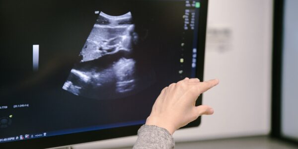
- Ultrasonography is an ultrasound based diagnostic imaging technique. It is mainly used to visualize internal body organs.
- A sound wave is typically produced by a piezoelectric transducer. A piezoelectric element produces a voltage across their two surfaces when deformed.
- Strong electrical pulses from the ultrasound machine make the transducer oscillate at desired frequency. The sound is focused to the concerned body part either by the shape of the transducer or by the process called beamforming.
- The returning reflections from the desired part of body make the transducer to vibrate and these vibrations are reconverted into electrical pulses.
- The ultrasonic scanner determines how long it took the echoes to return back to the transducer after the sound wave was sent. After this reception of echoes the ultrasonic scanner produces a digital image.
- In A-scan mode a single transducer fires sound pulses into the tissue and the echoes are analyzed.
- Strong echoes are marked with greater amplitude and weaker echoes are marked, with less amplitude in an oscilloscope. Hence it is also called Amplitude (A) mode.
- B-scan mode is called so because here the stronger echoes are marked with greater brightness.
- Here M-mode scan means motion scan .In T-M mode scanning a sequence of B-mode scans are moved rapidly to enable to measure the range of motion.
- Transcutaneous flow detectors are designed to use the Doppler Effect to detect the flow of blood in arteries close to the surface of the body.
- By exactly calculating the frequency shift caused due to Doppler Effect, we can indirectly convert it to the flow rate of blood.
- Ultrasonic techniques can be used to measure arterial blood pressure indirectly with the same method as used for flow detection.
- It has no long term side effects and comfortable and also images the muscles, bone surfaces, soft tissues etc well.
- The performance of sonographic system degrades when there is gas between the transducer and the organ of interest .It is due to the change in acoustic impedance.
- Medical ultrasonography is used in the study of many different systems such as cardiology, ophthalmology, neurology etc.
- Focused ultrasound can also be used to break up kidney stones.
- Low intensity ultrasound exposure can help in stimulating bone-growth and High Intensity Focused Ultrasound (HIFU) is used for the treatment of diseases such as cancer.
Author Bio: The Author of this article, Sreejith is writing articles on Ultrasonic Flow Meter Diagram and Electronics and Communications